Our Dermatology Team examines and treats a multitude of dermatologic disorders. In order to provide your pet the best care, our team uses the newest diagnostic tests and equipment including collection of a variety of skin samples (examined under microscope), skin biopsies, video otoscopy (including deep ear flushes and mass removal), advanced imaging (computed tomography), intradermal allergy testing for environmental allergies and CO2 (carbon dioxide) laser procedures of the skin. A brief description of these procedures is provided below, but please consult with one of our team members if more specific information is needed.
Cytology, Skin Scraping and Trichogram
Cytology is performed in almost every animal to look for any active infections that are present on the skin. These infections can look like other diseases and be a big cause of itching in our affected animals. Skin scraping is a common diagnostic tool used to evaluate for any parasite that may be present in the skin and affecting the animal. Trichograms are also performed to evaluate the hairs and look for any abnormalities or infectious agents that are affecting the hairs.
Skin Biopsies
Sometimes, we will need to take a small sample of skin from your pet and process that sample for microscopic evaluation by a pathologist to identify the underlying problem. This may be done with a punch biopsy to take a core of the tissue or surgical excision of an area with a scalpel blade or CO2 laser. Depending on the patient and location of the area, this can be done with either a local anesthetic, sedation or under general anesthesia. This diagnostic tool plays a critical part to establish a diagnose of autoimmune diseases, cancer (neoplasia), and inflammatory conditions.
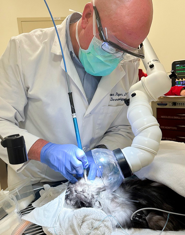
The ear is viewed through a video otoscope which provides a magnified and detailed view of the ear to look for normal structures as well as identify any abnormalities. The video otoscope is also used to perform deep ear flushes of the ear to remove debris from the ear and evaluate deeper within the ear canal. Abnormalities, such as a mass, a foreign body, or infection within the middle ear canal, can then be visualized and tools can be used through the ports within the video otoscope to remove these for further diagnostics, if necessary.
Advanced Imaging (Computed Tomography)
Advanced imaging can be performed of the entire head, with a focus on the ears in most cases. These images can help to identify the full extent of the disease present in the ear, including infection and masses within the middle ear that are not visible using our video otoscope.
Intradermal Allergy Testing (IDAT)
If the animal is diagnosed with environmental allergies, we perform IDAT to identify what environmental allergens they have an allergic response towards. IDAT is not used to diagnose environmental allergies but rather used to develop an allergen specific immunotherapy for that individual animal.
CO2 (Carbon Dioxide) Laser
CO2 laser is a common tool used to surgically remove masses that may not fit within a smaller punch biopsy. Additionally, some common and benign masses or inflammatory conditions are treated with just ablation (vaporization) to remove the affected areas and allow the skin to heal on its own. The CO2 laser is commonly used on skin as well as within the ear canal.
Clinical Trials
As part of the University and College’s goal of improving care for pets, our faculty and staff are frequently engaged in clinical trials to identify improved methods of diagnosing diseases or investigations on novel therapeutics to provide the best treatments to improve patient outcomes.
There are no current dermatology clinical trials at this time.